
The heartbeat is a vital physiological function driven by the cardiac conduction system, a specialized network of cells and tissues in the heart. This system initiates and coordinates the electrical signals responsible for the rhythmic contractions of the heart muscle. Here’s a detailed note on the elements of the cardiac conduction system and the heartbeat:
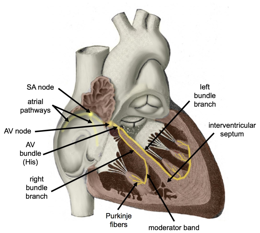
1. Sinoatrial (SA) Node:
Located in the right atrium near the opening of the superior vena cava.
Often referred to as the “natural pacemaker” of the heart,
Generates electrical impulses regularly (about 60–100 times per minute), initiating each heartbeat.
These impulses travel through the atria, causing them to contract and push blood into the ventricles.
2. Atria:
The electrical signals from the SA node stimulate the contraction of the atria, forcing blood into the ventricles.
Atrial contraction accounts for about 20–30% of the blood filling the ventricles.
3. Atrioventricular (AV) Node:
Located at the junction of the atria and ventricles in the interatrial septum.
serves as a relay station, slowing down the electrical impulses for a brief moment.
This delay allows the ventricles adequate time to fill with blood before they contract.
If the SA node fails, the AV node can take over the role of the pacemaker, but at a slower rate (about 40–60 times per minute).
4. Bundle of His (AV Bundle):
After passing through the AV node, the electrical signals travel down His bundle.
This bundle connects the atria and ventricles.
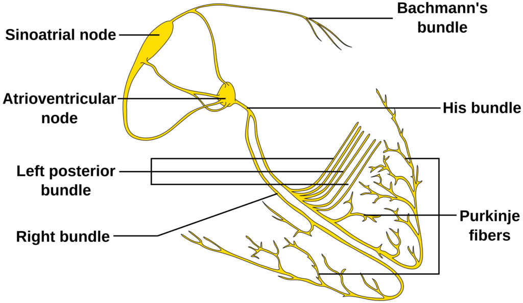
5. Right and Left Bundle Branches:
The bundle of His divides into right and left bundle branches, which extend down either side of the interventricular septum.
6. Purkinje fibers:
At the end of the bundle branches, the electrical impulses spread into a network of specialized fibers called Purkinje fibers.
These fibers are distributed throughout the ventricles.
They rapidly transmit electrical signals to the ventricular muscle cells, causing coordinated ventricular contraction.
7. Ventricles:
The electrical impulses from the Purkinje fibers trigger the ventricular muscle cells to contract.
Ventricular contraction forces blood out of the heart, with the left ventricle pumping oxygenated blood into the systemic circulation and the right ventricle pumping deoxygenated blood into the pulmonary circulation.