The osseous system, also known as the skeletal system, is an integral part of the human body that provides structure, support, and protection. It is a complex framework of bones, cartilage, and other connective tissues that serves several crucial functions in the body.
The axial skeleton is one of the two main divisions of the human skeleton, the other being the appendicular skeleton. It consists of the bones along the central axis of the body, which includes the skull, vertebral column, and ribcage. These bones have distinct structures and functions essential for various physiological processes and overall body support. Here is a detailed note on the structure and functions of the bones of the axial skeleton:
1. Skull:
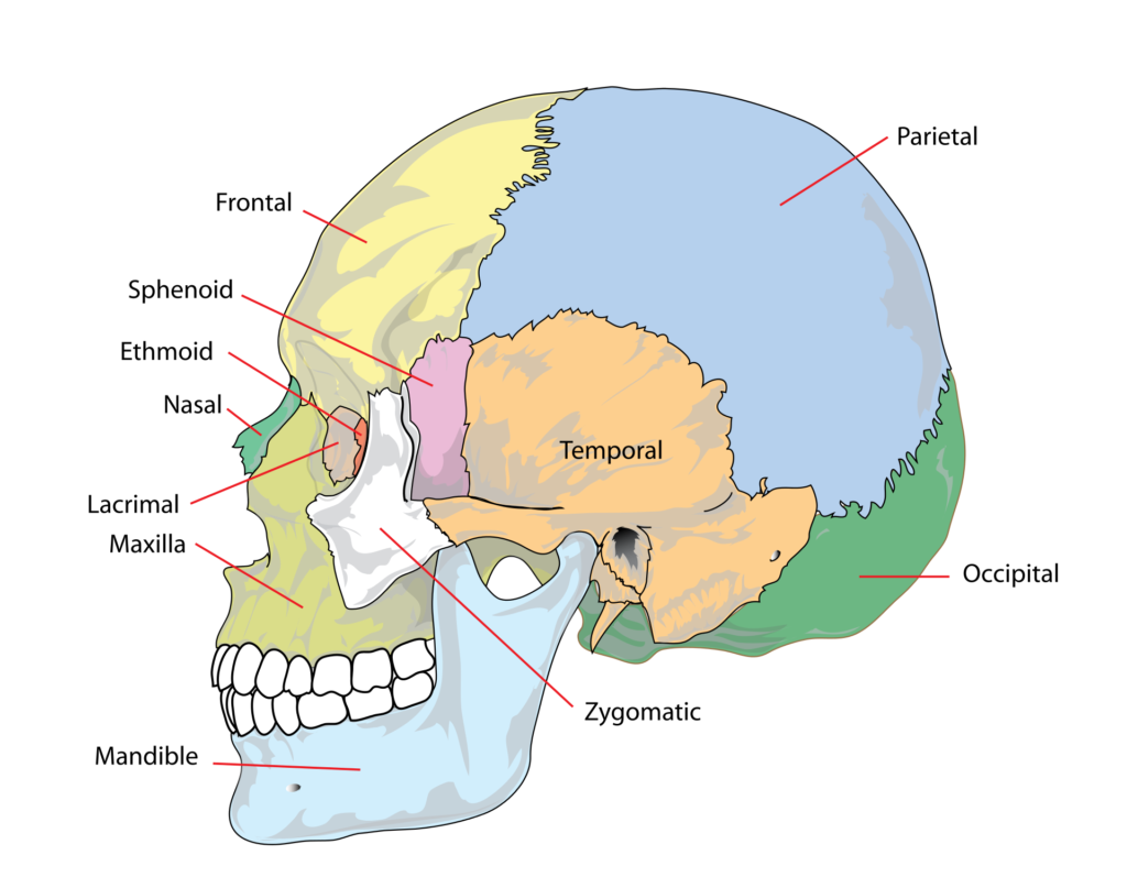
Cranium: The cranium is the part of the skull that surrounds and protects the brain. It consists of several flat, irregular bones, including the frontal bone (forehead), parietal bones (top and sides of the skull), temporal bones (sides of the head), and occipital bone (back of the head).
Facial Bones: The facial bones, such as the mandible (lower jaw), maxilla (upper jaw), and zygomatic bones (cheekbones), provide structure to the face and house the oral and nasal cavities. The nasal bones, nasal conchae, and others contribute to the nasal structure and the sinuses.
Functions: The skull protects the brain and sensory organs (eyes, ears, and nose). It also provides the framework for facial features, facilitates chewing, and supports various muscles of facial expression and mastication.
2. Vertebral Column (Spine):
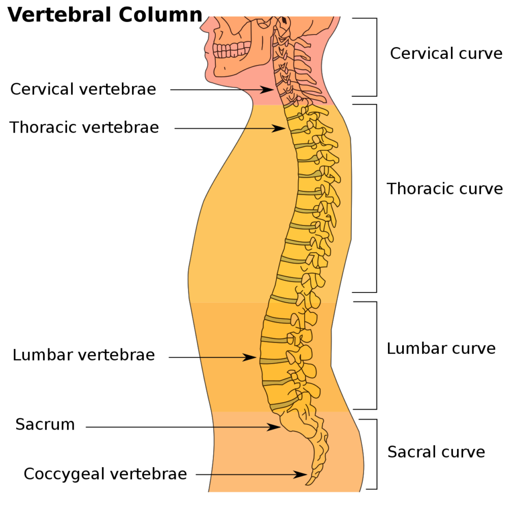
Vertebrae: The vertebral column consists of 33 vertebrae, which are divided into five regions: cervical (neck), thoracic (upper/mid-back), lumbar (lower back), sacral (pelvic), and coccygeal (tailbone). Each vertebra has a distinctive structure with intervertebral discs between them, made of cartilage.
Functions: The vertebral column supports the body’s weight and maintains an upright posture. It also protects the spinal cord, allowing for spinal cord function, and permits various degrees of flexibility and movement.
3. Ribcage (Thoracic Cage):
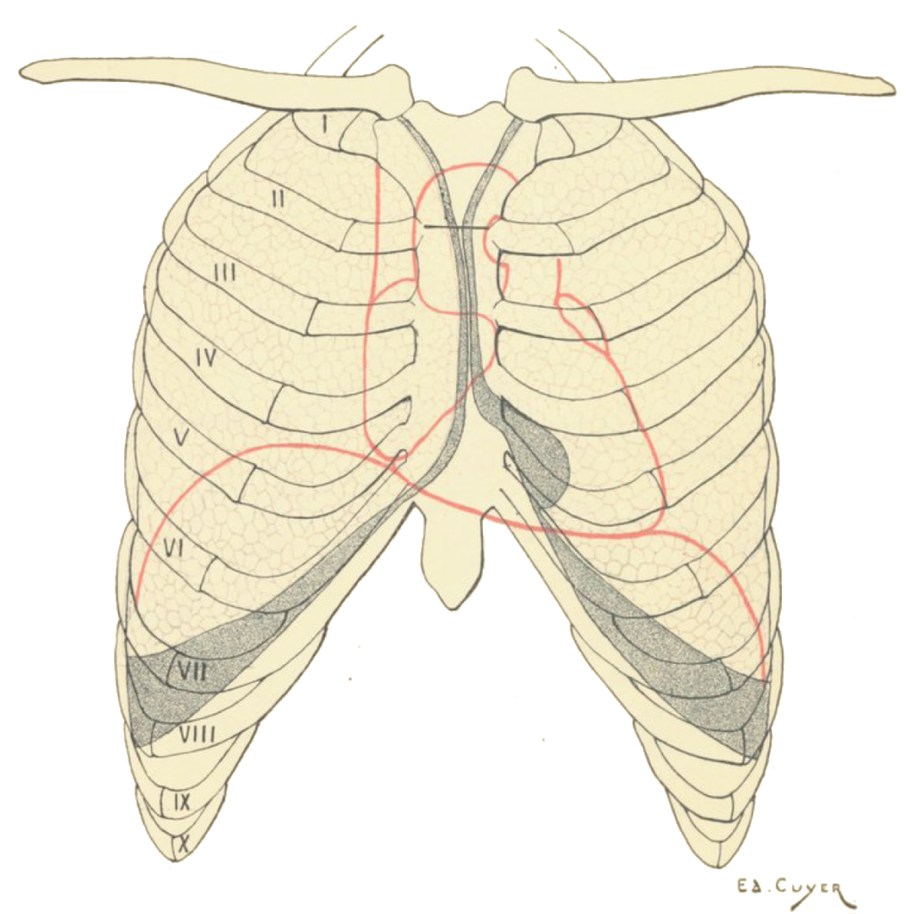
Ribs: The ribcage consists of 12 pairs of ribs that attach to the thoracic vertebrae. Seven pairs are true ribs (directly attach to the sternum via costal cartilage), and the remaining five pairs are false ribs (attach indirectly to the sternum or don’t attach at all).
Sternum (Breastbone): The sternum is a flat bone in the center of the chest that connects the ribcage. It consists of the manubrium, body, and xiphoid process.
Functions: The ribcage provides protection to the vital organs in the thoracic cavity, including the heart and lungs. It also plays a role in respiration by enabling the expansion and contraction of the thoracic cavity during breathing.
4. Hyoid Bone:
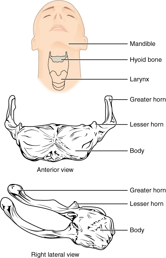
The hyoid bone is a U-shaped bone located in the neck, just below the mandible. It is unique because it doesn’t directly articulate with any other bone.
Function: The hyoid bone serves as an anchor point for various muscles involved in swallowing, speaking, and neck movement.
In summary, the axial skeleton consists of the bones of the skull, vertebral column, ribcage, and hyoid bone. Each of these bone structures has specific functions that are crucial for protecting vital organs, facilitating movement, and maintaining posture. Together, they provide essential support and protection for the body’s central axis and enable various physiological processes, including brain protection, spinal cord function, and respiration.
Appendicular skeleton system
The appendicular skeleton is one of the two main divisions of the human skeleton, with the other being the axial skeleton. It consists of the bones of the upper and lower limbs, as well as the girdles that attach these limbs to the axial skeleton. The appendicular skeleton plays a crucial role in body movement, support, and various functions. Here’s a detailed note on the structure and functions of the bones of the appendicular skeleton:
1. Pectoral (Shoulder) Girdle:
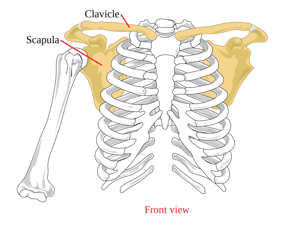
Clavicle (collarbone): The clavicle is a slender, S-shaped bone that connects the scapula to the sternum.
Scapula (Shoulder Blade): The scapula is a flat, triangular bone that attaches to the clavicle and provides attachment points for several muscles.
Functions: The pectoral girdle, along with the associated muscles, allows for the movement of the arms. It provides flexibility and stability for various arm movements.
2. Upper Limbs:
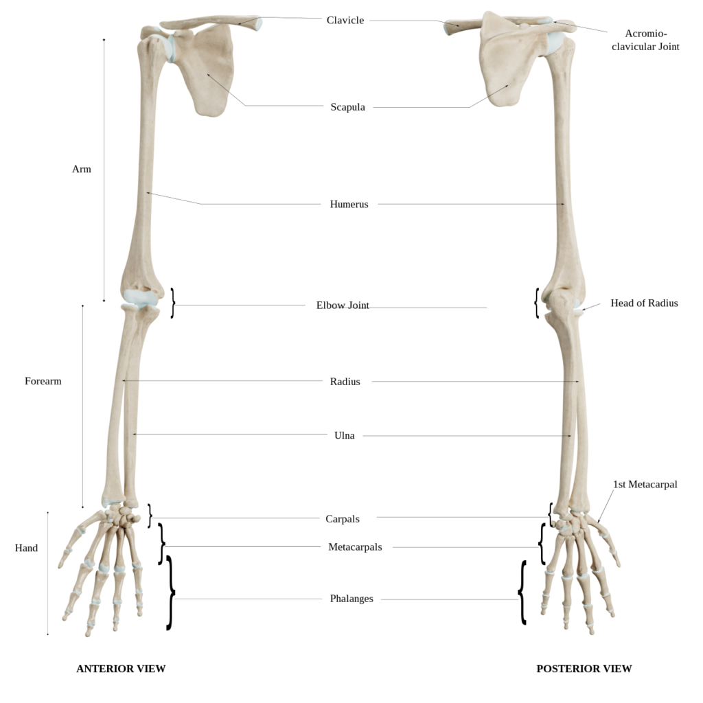
Humerus: The humerus is the bone of the upper arm, connecting the shoulder to the elbow.
Radius and Ulna: The radius and ulna are the bones of the forearm. The radius is on the thumb side, and the ulna is on the pinky side.
Carpal Bones: These are the bones of the wrist, including the scaphoid, lunate, triquetrum, and others.
Metacarpal Bones: These are the bones of the palm of the hand.
Phalanges: These are the finger bones, with three in each finger and two in the thumb.
Functions: The upper limbs and their bones enable a wide range of movements and functions, including reaching, grasping, manipulating objects, and interacting with the environment.
3. Pelvic (Hip) Girdle:
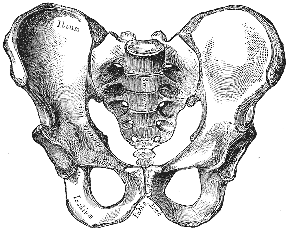
Ilium, Ischium, and Pubis: These three bones fuse to form the pelvic girdle or hip bones. The ilium is the large, flaring bone, the ischium forms the curved base, and the pubis is at the front.
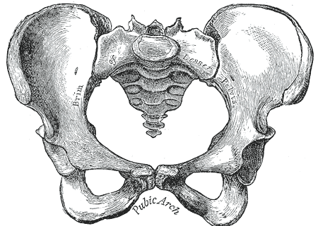
Functions: The pelvic girdle supports the weight of the body and protects internal reproductive and digestive organs. It also serves as an attachment point for muscles and ligaments involved in leg movement.
4. Lower Limbs:
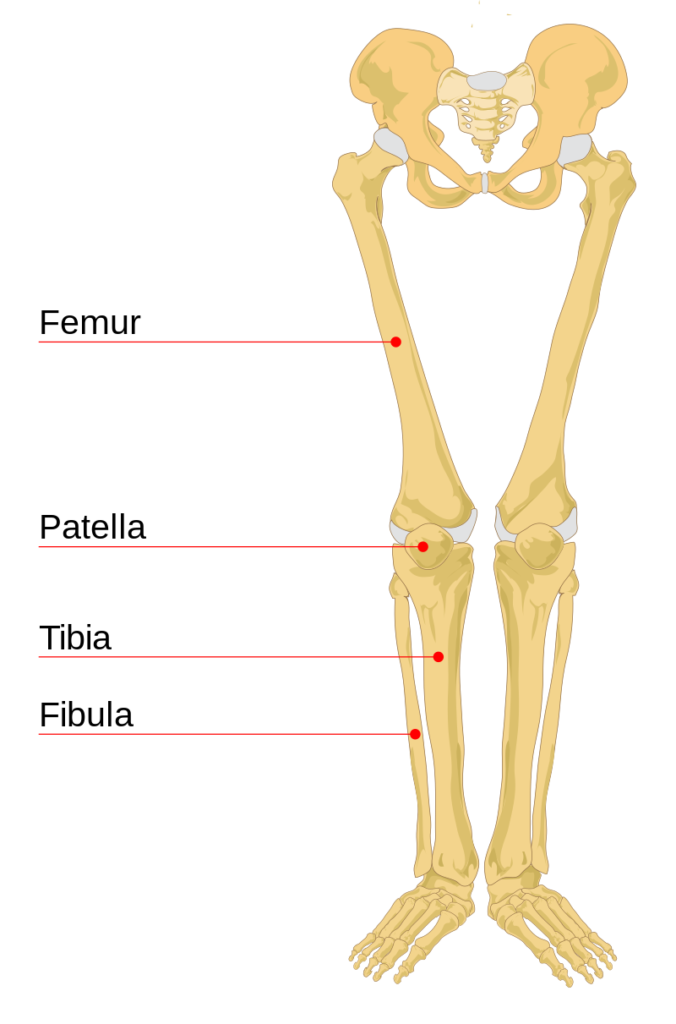
Femur: The femur is the thigh bone connecting the hip to the knee.
Tibia and Fibula: The tibia is the larger of the two leg bones and is situated on the inner side, while the fibula is thinner and located on the outer side.
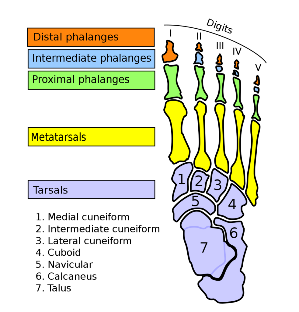
Tarsal Bones: These are the bones of the ankle, including the calcaneus (heel bone), talus, and others.
Metatarsal Bones: These are the bones of the foot’s arch.
Phalanges: These are the toe bones, similar to the phalanges in the fingers.
Functions: The lower limbs are responsible for weight-bearing, locomotion, and various physical activities such as walking, running, and jumping. They support the body’s weight and help maintain balance.