

The kidneys are vital organs responsible for maintaining fluid balance, electrolyte levels, and the removal of waste products from the bloodstream. Within the kidneys, nephrons are the functional units responsible for these processes. Let’s explore the anatomy of the kidneys and nephrons in detail:
Anatomy of the Kidneys
1. Location and General Structure
The kidneys are paired organs located in the posterior part of the abdominal cavity, on either side of the spine. Each kidney is roughly bean-shaped and about the size of a fist. The kidneys are retroperitoneal, meaning they are situated behind the peritoneum, the lining of the abdominal cavity. They are protected by the lower ribs and surrounded by a layer of adipose tissue that helps cushion and support them.
2. External Anatomy
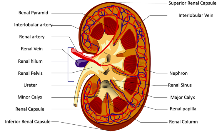
The kidney has a convex lateral border and a concave medial border. The renal hilum, located on the medial side, is a notch where the renal artery, renal vein, nerves, and ureter enter and exit the kidney. The renal capsule, a fibrous connective tissue layer, surrounds each kidney, providing structural support and protection.
3. Internal Anatomy
Each kidney is divided into two main regions: the outer renal cortex and the inner renal medulla. The renal cortex contains nephrons, renal corpuscles, and renal tubules, while the renal medulla consists of renal pyramids and renal columns. Renal pyramids are cone-shaped structures with their bases facing the cortex and their apexes, called renal papillae, projecting into the renal pelvis. Renal columns are extensions of cortical tissue that separate adjacent renal pyramids and help support the medullary tissue.
4. Blood Supply
The kidneys receive a large volume of blood flow, accounting for about 20-25% of the cardiac output. Blood enters each kidney through the renal artery, which branches into smaller arteries within the kidney. After filtration and waste removal in the nephrons, clean blood exits the kidneys through the renal vein and returns to the systemic circulation.
Anatomy of Nephrons
1. General Structure: Nephrons are the functional units of the kidneys responsible for filtering blood and producing urine. Each kidney contains millions of nephrons, which consist of a renal corpuscle and renal tubules. The renal corpuscle comprises the glomerulus, a tuft of capillaries, and the Bowman’s capsule, a cup-shaped structure surrounding the glomerulus.
2. Renal Tubules: The Bowman’s capsule leads into the renal tubules, which consist of the proximal convoluted tubule (PCT), loop of Henle, distal convoluted tubule (DCT), and collecting ducts. The PCT is located closest to the renal corpuscle and is responsible for reabsorbing most of the filtered water and essential solutes from the filtrate.
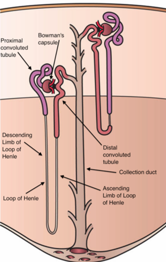
The loop of Henle descends into the renal medulla and consists of a descending and ascending limb, playing a crucial role in establishing and maintaining the osmotic gradient in the kidney. The DCT is responsible for further reabsorption and secretion of specific ions and substances, under the influence of hormones such as aldosterone and antidiuretic hormone (ADH). Collecting ducts receive filtrate from multiple nephrons and are responsible for final adjustments to urine volume and composition before it is excreted from the body.
3. Blood Filtration: Blood is filtered in the glomerulus under high pressure, allowing water, ions, and small molecules to pass through the filtration membrane into the Bowman’s capsule. Large proteins and blood cells are retained in the bloodstream due to the size-selective properties of the filtration membrane.
4. Urine Formation: Filtrate from the renal tubules undergoes processes of reabsorption and secretion to produce urine. Reabsorption involves the movement of water and solutes from the filtrate back into the bloodstream, while secretion involves the transfer of substances from the bloodstream into the filtrate for excretion. The composition of urine is finely regulated to maintain homeostasis by adjusting the reabsorption and secretion rates based on the body’s needs.
Understanding the intricate anatomy of the kidneys and nephrons is essential for comprehending their physiological functions in maintaining proper fluid balance, electrolyte levels, and waste elimination within the body. Dysfunction or damage to these structures can lead to various renal disorders and systemic health issues, highlighting the critical importance of kidney health for overall well-being.