

Cardiovascular system
The heart (Cardiovascular system) is a vital organ in the human body and is often described as a muscular pump responsible for circulating blood throughout the entire circulatory system. It plays a central role in maintaining life by ensuring the delivery of oxygen and nutrients to various tissues and organs while removing waste products. Here’s a general description of the heart:
Location: The heart resides in the thoracic cavity within the mediastinum—the central compartment between the lungs. It is slightly tilted to the left of the body’s midline. The base of the heart lies at the level of the second rib, while the apex, the pointed end, angles downward and to the left, resting near the fifth intercostal space. This strategic placement allows the heart to be protected by the rib cage while remaining accessible to major vessels.
Size: The heart is roughly the size of a closed fist and typically weighs between 200 to 425 grams (approximately 7 to 15 ounces). However, its size can vary depending on factors such as an individual’s age, sex, body size, and physical condition. Athletes, for example, may have slightly larger hearts due to increased workload and endurance training.
Shape: The shape of the heart resembles a cone, with a broader base and a pointed apex. This conical structure supports efficient contraction and ejection of blood into major arteries. The heart’s muscular design plays a crucial role in maintaining blood circulation. It is made primarily of cardiac muscle, a type of involuntary, striated muscle known for its endurance and rhythmic contractility.
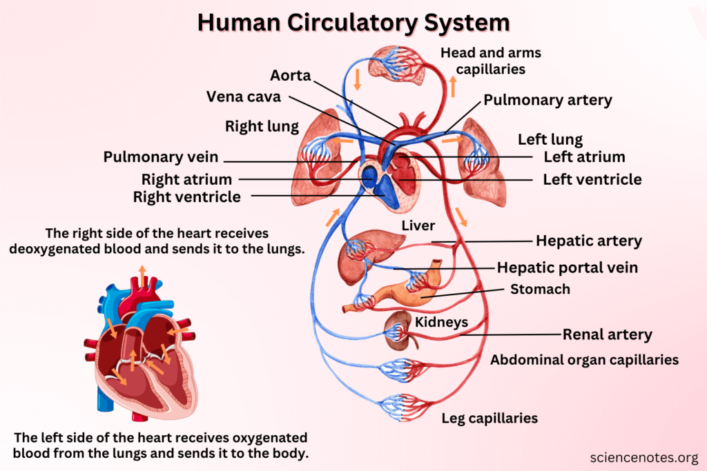
Chambers: The heart consists of four chambers: two upper chambers known as atria and two lower chambers known as ventricles. The right atrium receives deoxygenated blood from the body through the superior and inferior vena cava, while the left atrium receives oxygenated blood from the lungs via the pulmonary veins. The right ventricle pumps deoxygenated blood to the lungs through the pulmonary artery, and the left ventricle, which has the thickest walls, pumps oxygenated blood to the entire body through the aorta.
Valves: To ensure the unidirectional flow of blood, the heart is equipped with four valves:
The tricuspid valve sits between the right atrium and right ventricle.
The pulmonary valve is located between the right ventricle and the pulmonary artery.
The mitral (or bicuspid) valve lies between the left atrium and left ventricle.
The aortic valve is found between the left ventricle and the aorta.
These valves open and close in coordination with each heartbeat, preventing backflow and ensuring the efficient circulation of blood.
Blood Supply: The heart is nourished by its own blood supply, provided by the coronary arteries. These arteries branch from the base of the aorta and encircle the heart muscle, delivering oxygen and nutrients essential for its continuous activity. Blockage or damage to these arteries can result in ischemia or myocardial infarction (heart attack).
Electrical System: Underlying the heart’s rhythmic beating is its electrical conduction system, which orchestrates the timing and force of each contraction. This system includes the sinoatrial (SA) node, often referred to as the natural pacemaker, which initiates electrical impulses. The impulse then travels to the atrioventricular (AV) node, the bundle of His, and eventually through the Purkinje fibers, ensuring coordinated contraction of the atria and ventricles.
Layers of heart
The walls of the heart are composed of three primary layers, each contributing to its function:
Endocardium: The endocardium is the innermost layer lining the heart’s chambers and valves. It consists of a thin, smooth endothelial lining that minimizes resistance to blood flow and reduces friction. It also plays a role in the heart’s electrical conduction, as it contains specialized cells that facilitate impulse transmission.
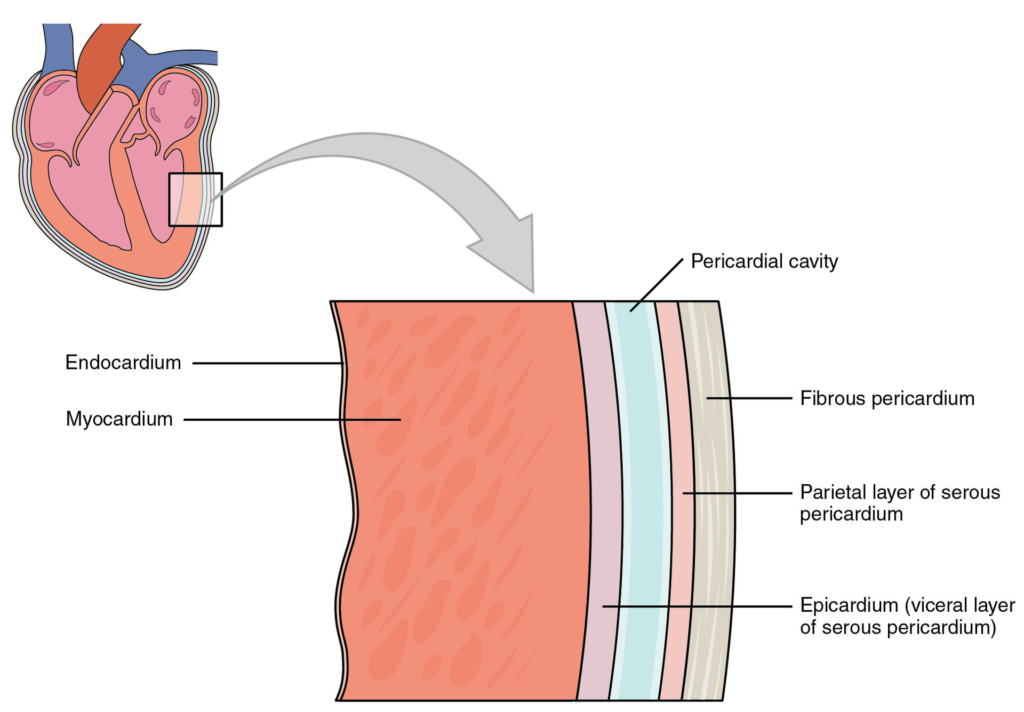
Myocardium: The myocardium forms the thick, muscular middle layer and is responsible for the heart’s contractile power. Composed of cardiac muscle fibers, the myocardium contracts rhythmically and involuntarily to pump blood throughout the body. This layer is particularly thick in the left ventricle, which must generate significant force to propel blood into the systemic circulation.
Epicardium: The epicardium is the outermost layer of the heart wall and is also considered the visceral layer of the serous pericardium. It serves a protective function and contains connective tissue, blood vessels, nerves, and a layer of fat that cushions the heart. The epicardium also helps in secreting a lubricating fluid that reduces friction between the heart and surrounding pericardial layers during contraction.
Together, these structural components enable the heart to fulfill its vital role in sustaining life by maintaining continuous and controlled blood flow to all parts of the body.
Functions of the Cardiovascular System
Transport of Respiratory Gases: One of the most vital roles of the cardiovascular system is the transportation of respiratory gases. Oxygen, which is inhaled into the lungs, is picked up by red blood cells and carried through the bloodstream to various tissues and organs where it is needed for cellular respiration. At the same time, carbon dioxide—a waste product of metabolism—is collected from the tissues and transported back to the lungs to be exhaled.
Distribution of Nutrients: After digestion, essential nutrients such as glucose, amino acids, fatty acids, vitamins, and minerals are absorbed into the blood from the gastrointestinal tract. The cardiovascular system is responsible for distributing these nutrients to all parts of the body where they are required for energy production, growth, and repair.
Removal of Metabolic Waste Products: The cardiovascular system plays a crucial role in waste elimination by transporting metabolic by-products such as urea, creatinine, and carbon dioxide from tissues to organs like the kidneys, lungs, and liver. These organs then process and eliminate the waste through urine, feces, or exhaled air.
Transport of Hormones and Other Signaling Molecules: Hormones released by endocrine glands such as the pituitary, thyroid, and adrenal glands are circulated throughout the body via the bloodstream. This ensures that chemical messages are delivered efficiently to target tissues and organs, allowing for the coordination of complex bodily functions like growth, metabolism, reproduction, and stress response.
Regulation of Body Temperature: Blood flow plays an essential role in thermoregulation. When body temperature rises, blood vessels near the surface of the skin dilate to release heat. Conversely, when it’s cold, these vessels constrict to conserve heat. Thus, the cardiovascular system helps maintain a stable internal temperature.
Maintenance of pH and Electrolyte Balance: The cardiovascular system helps to stabilize the body’s pH levels through buffer systems in the blood, such as bicarbonate, and maintains the correct concentration of electrolytes like sodium, potassium, calcium, and chloride. This balance is critical for normal nerve function, muscle contraction, and overall metabolic processes.
Protection Against Disease and Blood Loss: The cardiovascular system is also a defense mechanism. It includes components of the immune system such as white blood cells, which help detect and destroy pathogens. Antibodies and other immune proteins in the blood provide immunity. Additionally, platelets and clotting factors help form blood clots to prevent excessive blood loss in case of injury.
 Join Our Telegram Channel
2 thoughts on “Cardiovascular system: Definition, Structure, Functions”