

Central Nervous System
The central nervous system (CNS) comprises the brain and spinal cord, encased in protective layers and filled with cerebrospinal fluid (CSF). Understanding the anatomy and functions of the meninges, ventricles of the brain, and CSF is crucial for comprehending CNS health, pathology, and therapeutic interventions.

1. Meninges
The meninges are three protective layers surrounding the brain and spinal cord. They are (from outermost to innermost):
Dura Mater: This tough, fibrous membrane is the outermost layer. It consists of two layers: the outer periosteal layer, which attaches to the inner surface of the skull, and the inner meningeal layer, which forms the dural folds and supports venous sinuses.
Arachnoid Mater: This delicate, web-like membrane lies beneath the dura mater. The subarachnoid space separates it from the pia mater and contains trabeculae, which attach it to the pia mater. The arachnoid mater also has extensions called arachnoid villi, which protrude into the venous sinuses and are involved in CSF absorption.
Pia Mater: This innermost layer is a thin, delicate membrane that closely adheres to the brain and spinal cord, following their contours.
The meninges protect the CNS from mechanical trauma, provide stability and support, and participate in the production and circulation of CSF.
2. Ventricles of the Brain
The ventricular system consists of interconnected cavities within the brain filled with CSF. There are four ventricles:
Lateral Ventricles (First and Second Ventricles): These are the largest ventricles and are located in the cerebral hemispheres. Each lateral ventricle consists of a body, anterior horn, posterior horn, and inferior horn.
Third Ventricle: Located in the diencephalon, between the two halves of the thalamus. It communicates with the lateral ventricles via the interventricular foramen (of Monro) and with the fourth ventricle via the cerebral aqueduct (of Sylvius).
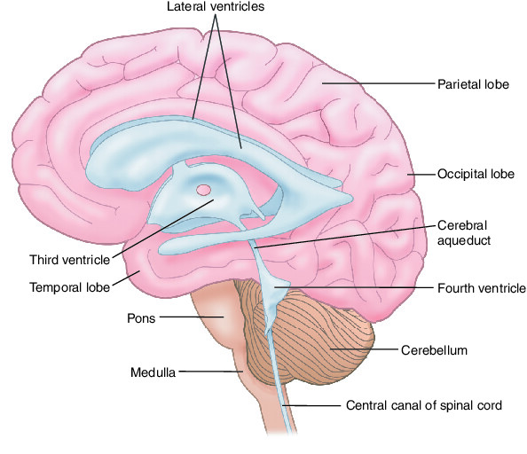
Fourth Ventricle: Situated in the brainstem, between the pons and the cerebellum. It communicates with the third ventricle via the cerebral aqueduct and the subarachnoid space surrounding the brain and spinal cord via three openings: the median aperture (of Magendie) and two lateral apertures (of Luschka).
The ventricles contain choroid plexuses, specialized structures that produce CSF. CSF flows from the lateral ventricles through the interventricular foramina into the third ventricle, then through the cerebral aqueduct into the fourth ventricle, and finally into the subarachnoid space.
3. Cerebrospinal Fluid (CSF)
CSF is a clear, colorless fluid that fills the ventricles of the brain, the subarachnoid space surrounding the brain and spinal cord, and the central canal of the spinal cord. It serves several vital functions:
Buoyancy: CSF supports and cushions the brain, reducing its effective weight by about 95%, thereby preventing mechanical injury.
Protection: CSF provides a protective cushion against physical trauma and helps to remove waste products from the CNS.
Chemical Stability: CSF regulates the chemical environment of the CNS by transporting nutrients and removing metabolic waste products.
Volume Transmission: CSF acts as a medium for the distribution of neuromodulators and signaling molecules throughout the CNS.
CSF is continuously produced by the choroid plexuses at a rate of about 500 mL per day, with the total volume of CSF in the CNS typically around 125-150 mL. It is absorbed primarily by arachnoid villi protruding into the venous sinuses, although some absorption also occurs through the meningeal lymphatics.
Clinical Relevance:
Disorders affecting the meninges, ventricles, or CSF can have serious consequences for CNS function and health. Examples include:
Meningitis: Inflammation of the meninges, often caused by infection, leading to symptoms such as headache, fever, stiff neck, and altered mental status.
Hydrocephalus: Accumulation of excess CSF within the ventricles, leading to increased intracranial pressure, enlarged ventricles, and compression of brain tissue. It can result from obstruction of CSF flow, overproduction of CSF, or impaired CSF absorption.
Subarachnoid Hemorrhage: Bleeding into the subarachnoid space, often due to rupture of a cerebral aneurysm, leading to sudden, severe headache (“thunderclap headache”), nausea, vomiting, and neurological deficits.
Understanding the anatomy and physiology of the meninges, ventricles, and CSF is essential for diagnosing and managing these and other CNS disorders effectively. Advanced imaging techniques such as MRI and CT scans are crucial in visualizing these structures and evaluating abnormalities.