

Circulation of Blood in the Heart: Blood circulation in the heart involves a complex and highly regulated process that allows blood to move efficiently, delivering oxygen and nutrients to body tissues while removing waste products. The heart plays a central role in this process, pumping blood to the lungs for oxygenation and the rest of the body’s organs and tissues. Here is a detailed note on the circulation of blood in the heart:
Check this post: Anatomy of the heart
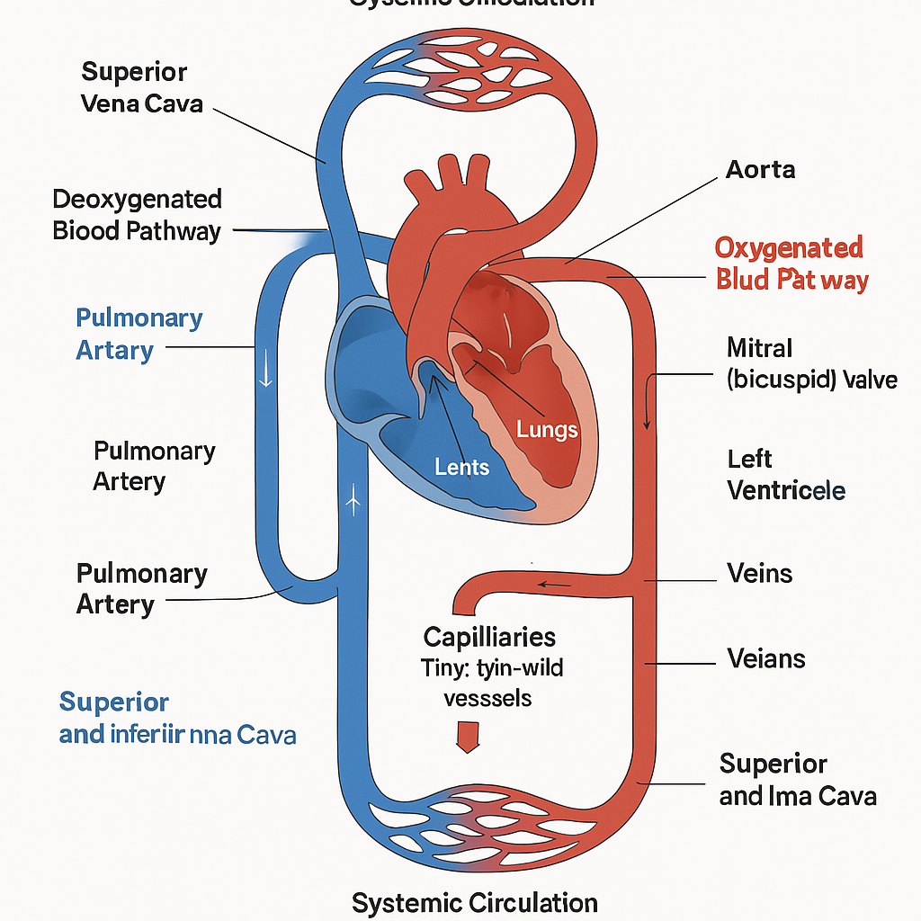
1. Deoxygenated Blood Pathway
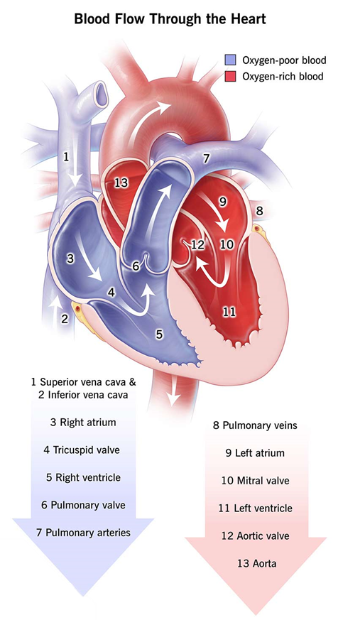
a. Superior and Inferior Vena Cava: Deoxygenated blood, which has delivered oxygen to the body tissues and picked up carbon dioxide and other waste products, returns to the heart through two large veins. The superior vena cava brings blood from the upper parts of the body such as the head, neck, arms, and chest, while the inferior vena cava collects blood from the lower regions, including the legs, abdomen, and pelvis. These two major vessels serve as the main entry routes for deoxygenated blood into the heart.
b. Right Atrium: The blood enters the right atrium, which is the upper right chamber of the heart. This chamber acts as a receiving station, temporarily holding the deoxygenated blood before it is sent into the right ventricle. As the atrium fills with blood, it prepares to contract and push the blood forward into the next chamber of the heart.
c. Tricuspid Valve: When the right atrium contracts, the blood is forced through the tricuspid valve, which is a one-way valve located between the right atrium and the right ventricle. This valve ensures that blood flows in a forward direction and prevents any backflow into the atrium during the next phase of the cardiac cycle. The tricuspid valve opens under pressure and allows the blood to move into the right ventricle.
d. Right Ventricle: The right ventricle, which is the lower right chamber of the heart, receives the blood from the atrium and prepares to send it to the lungs. When the ventricle contracts, it generates enough pressure to pump the blood through the pulmonary valve and into the pulmonary artery. This marks the beginning of the blood’s journey to the lungs for oxygenation.
2. Pulmonary Circulation:
a. Pulmonary Artery: The pulmonary artery is the only artery in the body that carries deoxygenated blood. It transports the blood from the right ventricle to the lungs. This artery branches into the left and right pulmonary arteries, which lead to the corresponding lungs. The movement of blood through the pulmonary artery is essential for the removal of carbon dioxide and the uptake of oxygen in the lungs.
b. Lungs: In the lungs, the blood flows through a network of tiny capillaries surrounding the alveoli (air sacs). Here, a vital gas exchange takes place: carbon dioxide diffuses out of the blood into the alveoli to be exhaled, while oxygen from the inhaled air diffuses into the blood. As a result, the blood becomes oxygen-rich and is now ready to be returned to the heart to be pumped throughout the body.
2. Oxygenated Blood Pathway
a. Pulmonary Veins: After the blood has been oxygenated in the lungs, it travels back to the heart through the pulmonary veins. These are the only veins in the human body that carry oxygen-rich blood. There are four pulmonary veins—two from each lung—and they transport the oxygenated blood directly into the left atrium of the heart. This is a crucial part of the circulation as it marks the transition from pulmonary to systemic circulation.
b. Left Atrium: The left atrium is the upper left chamber of the heart. It receives freshly oxygenated blood from the pulmonary veins. This chamber acts as a temporary holding area for the blood before it moves into the left ventricle. As it fills, the atrium prepares to contract, which will push the blood into the next chamber through a specialized valve.
c. Mitral (bicuspid) Valve: When the left atrium contracts, the oxygen-rich blood is pushed through the mitral valve (also known as the bicuspid valve) into the left ventricle. This valve is located between the left atrium and the left ventricle and has two cusps or flaps. It functions to maintain one-way blood flow and prevents the backflow of blood into the left atrium during ventricular contraction.
d. Left Ventricle: The left ventricle is the lower left chamber of the heart and is the thickest and most muscular of all four chambers. Its powerful contractions are responsible for pumping oxygenated blood into the aorta, the largest artery in the body. From the aorta, the oxygen-rich blood is distributed through the systemic circulation to all parts of the body, supplying organs and tissues with the oxygen and nutrients they need for proper function.
3. Systemic Circulation
a. Aorta: The aorta is the largest and most important artery in the human body. It originates from the left ventricle of the heart and serves as the primary high-pressure conduit for oxygenated blood. When the left ventricle contracts, it sends a surge of oxygen-rich blood into the aorta. From there, the blood is directed into multiple branches that extend upward to the head and arms and downward to supply the rest of the body.
b. Arteries: As the blood flows away from the heart through the aorta, it is distributed into a vast network of arteries. These arteries progressively branch into smaller vessels, each one supplying oxygenated blood to specific tissues, organs, and cells throughout the body. The arterial system ensures that every part of the body receives a steady supply of oxygen and nutrients essential for cellular metabolism and function.
c. Capillaries: The capillaries are the smallest and most delicate blood vessels. They form an extensive network that connects the ends of arteries (arterioles) to the beginnings of veins (venules). Within the capillary beds, oxygen and nutrients are exchanged with the body’s cells. At the same time, waste products like carbon dioxide and metabolic byproducts move from the cells into the capillaries to be carried away. This is the critical site of nutrient delivery and waste removal at the cellular level.
d. Veins: Once the exchange in the capillaries is complete, the blood—now deoxygenated—begins its return journey to the heart. It is collected by small veins known as venules, which gradually merge into larger veins. These veins act as low-pressure vessels that transport the deoxygenated blood back toward the heart, aided by muscle contractions and one-way valves that prevent backflow.
e. Superior and Inferior Vena Cava: Finally, all the deoxygenated blood from the body is funneled into two major veins: the superior vena cava, which gathers blood from the head, neck, arms, and upper chest, and the inferior vena cava, which collects blood from the lower limbs, abdomen, and pelvis. Both of these large veins empty the deoxygenated blood into the right atrium of the heart, thereby completing the systemic circulation loop and preparing the blood to re-enter the pulmonary circuit for reoxygenation.