
Reversible cell injury occurs when cells experience extreme stress that they cannot immediately adapt to but can recover from if the stress is removed. This type of injury leads to cellular and morphological changes that are reversible. There are three primary mechanisms through which reversible cell injury can occur:
1. Depletion of ATP Resources
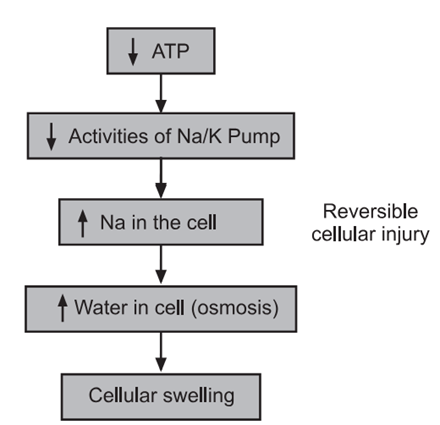
ATP Depletion: Reduced oxidative phosphorylation leads to lower ATP levels, which are crucial for various cellular processes, including membrane transport, protein synthesis, lipogenesis, and phospholipid turnover. When ATP levels drop to 5-10% of normal, critical cellular functions are impaired. The sodium-potassium pump fails, causing sodium to accumulate inside the cell and potassium to exit, leading to water influx, cell swelling, and endoplasmic reticulum dilation. Under ischemic conditions, cells switch to glycolysis for ATP production, depleting glycogen stores and accumulating lactic acid and inorganic phosphates, which lower intracellular pH and reduce enzyme activity.
2. Damage to Mitochondria
Mitochondrial Damage: Mitochondria, essential for ATP production, are susceptible to injury from increased cytosolic Ca²⁺, reactive oxygen species (ROS), and oxygen deprivation. Damage can lead to the formation of mitochondrial permeability transition pores, loss of membrane potential, and impaired oxidative phosphorylation, culminating in necrosis. Additionally, damaged mitochondria can release proteins like cytochrome C into the cytosol, triggering apoptosis. Cytochrome C is crucial for energy production within mitochondria but signals cell death when released.
3. Influx of Calcium
Calcium Influx: Failure of the Ca²⁺ pump results in harmful Ca²⁺ accumulation, disrupting protein synthesis by detaching ribosomes from the rough endoplasmic reticulum and breaking down polysomes into monosomes. This leads to reduced protein synthesis and ultimately to irreversible damage to mitochondrial and lysosomal membranes, causing necrosis. Misfolded proteins due to stress or damage can trigger the unfolded protein response, leading to further cell injury or death.
Additional Mechanisms Involved in Reversible Cell Injury
1. Defects in Membrane Permeability: Early loss of selective membrane permeability often leads to overt membrane damage due to ischemia, toxins, and physical or chemical agents.
2. Decreased Phospholipid Synthesis: Lower ATP levels reduce energy-dependent enzymatic activities, affecting phospholipid synthesis in cellular membranes, including mitochondria, exacerbating ATP loss.
3. Increased Phospholipid Breakdown: Severe cell injury activates endogenous phospholipases due to elevated cytosolic Ca²⁺, increasing membrane phospholipid degradation.
4. ROS: Reactive oxygen species damage cell membranes through lipid peroxidation.
5. Cytoskeletal Abnormalities: Increased cytosolic Ca²⁺ activates proteases that damage the cytoskeleton, which connects the plasma membrane to the cell interior.
6. Lipid Breakdown Products: Catabolic products like free fatty acids and lysophospholipids accumulate in injured cells, disrupting membranes through detergent effects or insertion into the lipid bilayer, altering permeability and electrophysiologic properties.
Sites of Membrane Damage
1. Mitochondrial Membrane Damage: Leads to decreased ATP production and release of apoptosis-inducing proteins.
2. Plasma Membrane Damage: Results in loss of osmotic balance, influx of fluids and ions, and leakage of vital metabolites, further depleting energy stores.
3. Lysosomal Membrane Damage: Causes leakage of enzymes into the cytoplasm, activating hydrolases that digest cell components, leading to necrosis.
Damage to DNA and Proteins
Cells attempt to repair DNA damage, but if damage is irreparable (e.g., due to radiation or oxidative stress), apoptosis is initiated. Similarly, improperly folded proteins from inherited mutations or external triggers, like free radicals, can cause apoptosis.
Reduced Protein Synthesis
Continued hypoxia causes swelling of endoplasmic reticulum and Golgi apparatus membranes, detaching ribosomes and degrading polysomes to monosomes, inactivating ribosome function and reducing protein synthesis. Withdrawal of acute stress at this stage can restore the cell to its normal state.