

Anatomy of the Urinary System
The urinary system, also known as the renal system, is responsible for the production, storage, and elimination of urine, which is the primary method by which the body rids itself of waste products and regulates various metabolic processes. The anatomy of the urinary tract encompasses a complex network of organs and structures that work together seamlessly to maintain proper fluid balance, electrolyte levels, and blood pressure within the body. Let’s delve into the detailed anatomy of the urinary tract:
1. Kidneys
The kidneys are bean-shaped organs located in the posterior part of the abdominal cavity, just below the diaphragm and on either side of the spine. Each kidney is approximately the size of a fist and is composed of millions of functional units called nephrons. Nephrons are the microscopic structural and functional units responsible for the filtration of blood and the formation of urine. The renal cortex is the outer layer of the kidney, while the renal medulla lies beneath it. Blood is supplied to the kidneys through the renal arteries, and filtered blood leaves the kidneys through the renal veins.
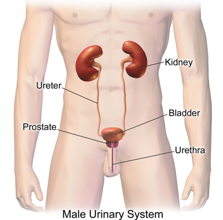
2. Ureters
The ureters are muscular tubes that connect the kidneys to the urinary bladder. There are two ureters, one arising from each kidney, and they transport urine from the renal pelvis to the bladder. Peristaltic contractions of smooth muscle in the ureter walls propel urine downward toward the bladder. The ureters enter the bladder obliquely, which helps prevent the backflow of urine into the kidneys.
3. Urinary Bladder
The urinary bladder is a hollow, muscular organ situated in the pelvis, posterior to the pubic symphysis. Its primary function is to store urine until it is expelled from the body during urination. The bladder has a highly distensible wall that allows it to expand as it fills with urine. It is composed of smooth muscle known as the detrusor muscle, which contracts during urination to expel urine through the urethra. The trigone is a triangular area within the bladder formed by the openings of the ureters and the urethra
4. Urethra
The urethra is a tubular structure that extends from the urinary bladder to the external urinary meatus, through which urine is expelled from the body. In males, the urethra is longer and traverses the prostate gland and the penis before opening at the tip of the penis. In females, the urethra is shorter and opens into the vestibule, located between the clitoris and the vaginal opening. The urethral sphincters, both internal and external, help control the flow of urine out of the bladder.
5. Accessory Organs:
The urinary system also includes several accessory organs that play supportive roles in the production and elimination of urine. These include the renal pelvis, which collects urine from the nephrons and funnels it into the ureters, and the urethral glands, which secrete mucus into the urethra to facilitate the passage of urine. Additionally, the adrenal glands, situated atop each kidney, produce hormones that regulate blood pressure, electrolyte balance, and fluid volume, all of which indirectly influence urinary function.
Understanding the detailed anatomy of the urinary tract is crucial for comprehending its physiological functions and the pathophysiology of various urinary disorders and diseases. Dysfunction or impairment of any part of the urinary system can lead to significant health problems, underscoring the importance of maintaining its proper structure and function.